IVUS and OCT Guided Coronary Intervention
IVUS and OCT Guided Coronary Intervention
Modern coronary intervention increasingly relies on advanced imaging techniques to understand the coronary arteries in greater detail. IVUS (Intravascular Ultrasound) and OCT (Optical Coherence Tomography) are specialised intravascular imaging technologies that provide detailed information about the inside of coronary arteries and help guide selected coronary interventions.
Dr. Pradeep Kumar Bansal, Chief Interventional Cardiologist and Heart Failure in Jaipur, has expertise in advanced interventional cardiology and provides individualised treatment planning for patients with coronary artery disease. IVUS and OCT can be used as part of a detailed assessment and stenting strategy when clinically appropriate.
What Are IVUS and OCT?
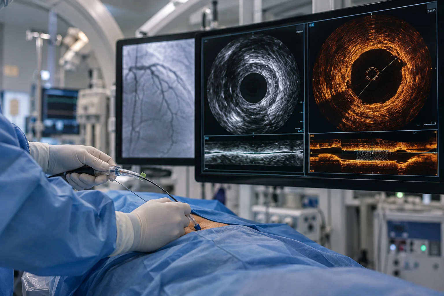
IVUS and OCT are intravascular imaging techniques used during coronary intervention to obtain detailed images from inside the blood vessel.
A conventional coronary angiogram provides a two-dimensional view of the coronary artery. Intravascular imaging can provide additional information about the vessel wall, plaque, narrowing and stent placement.
IVUS – Intravascular Ultrasound
IVUS uses ultrasound technology to create detailed images from within the coronary artery. It can help the interventional cardiologist assess the size and structure of the vessel and understand the characteristics of a coronary lesion.
OCT – Optical Coherence Tomography
OCT uses light-based imaging technology to provide high-resolution images of the inside of the coronary artery. Its detailed imaging capability can help evaluate selected coronary lesions and assess stent positioning and expansion.
Both technologies have an important role in selected cases, although the choice between IVUS and OCT depends on the patient's coronary anatomy, clinical situation and procedural requirements.
Why Is Intravascular Imaging Important?
Coronary angiography is an essential tool for diagnosing coronary artery disease, but certain details may not always be fully visible on angiography alone.
IVUS and OCT can provide additional information regarding:
- Vessel size and dimensions
- Plaque characteristics
- Extent of coronary narrowing
- Lesion morphology
- Stent positioning
- Stent expansion
- Areas requiring further optimisation
This additional information can help the interventional cardiologist make more informed decisions during selected coronary procedures.
IVUS-Guided Coronary Intervention
IVUS can be used during coronary intervention to assess the coronary artery from inside the vessel.
The imaging information may help the cardiologist understand the dimensions of the artery and characteristics of the lesion before selecting and positioning a stent.
After stent placement, IVUS can also be used to assess the result and determine whether further optimisation may be appropriate.
This makes IVUS particularly valuable when detailed vessel assessment is required during complex or technically challenging coronary interventions.
OCT-Guided Coronary Intervention
OCT provides high-resolution images of the coronary artery and can offer detailed visualisation of selected lesions and stents.
During an OCT-guided intervention, imaging can help assess the lesion before treatment and evaluate the stent after deployment.
The detailed images may assist the cardiologist in identifying areas where additional optimisation may be required, depending on the clinical circumstances.
IVUS vs OCT: Understanding the Difference
Although both IVUS and OCT are intravascular imaging technologies, they use different imaging principles and have different characteristics.
IVUS uses ultrasound and can provide information about the vessel structure and dimensions.
OCT uses light-based imaging and provides very high-resolution images of the vessel and stent.
The appropriate imaging method is selected according to the patient's coronary anatomy, lesion characteristics, procedural requirements and clinical judgement.
Who May Benefit From IVUS & OCT Guided Intervention?
Intravascular imaging may be considered for selected patients undergoing coronary intervention, particularly when detailed information is required beyond conventional angiography.
It may be considered in situations involving:
- Complex coronary lesions
- Uncertain vessel dimensions
- Detailed assessment of coronary plaque
- Selected challenging stenting procedures
- Assessment of stent expansion and positioning
- Need for procedural optimisation
Not every coronary intervention requires IVUS or OCT. The cardiologist determines whether intravascular imaging is appropriate based on the individual patient's condition.
How Is IVUS & OCT Guided Coronary Intervention Performed?
IVUS or OCT imaging is generally performed during a coronary catheter-based procedure.
A specialised imaging catheter or guidewire is carefully introduced into the coronary artery. Images are then obtained from within the vessel and assessed by the interventional cardiologist.
The findings can be used to support different stages of the procedure, including:
- Assessment of the coronary lesion
- Evaluation of vessel dimensions
- Planning of the intervention
- Guidance during stent placement
- Assessment of the treated area
- Optimisation of the final result when required
The exact procedure depends on the patient's coronary anatomy and the type of intervention being performed.
Role of IVUS and OCT in Stent Optimisation
Correct stent placement and expansion are important aspects of coronary intervention. In selected cases, intravascular imaging can provide detailed information that helps the cardiologist evaluate the treated segment.
Imaging may help identify areas where the stent has not achieved the desired expansion or positioning and may support further optimisation when clinically required.
This imaging-guided approach can be particularly useful when the coronary anatomy is complex or when angiography alone does not provide sufficient information.
Benefits of IVUS & OCT Guided Coronary Intervention
The potential advantages of intravascular imaging include:
Detailed Coronary Assessment
IVUS and OCT provide images from inside the coronary artery, offering information that complements angiography.
Better Understanding of Lesions
The imaging findings can help the cardiologist assess the characteristics and dimensions of selected coronary lesions.
Individualised Stent Planning
Information about vessel size and lesion characteristics can support more tailored stent selection and placement.
Assessment of Stent Results
Following stent deployment, imaging can help evaluate positioning and expansion.
Procedural Optimisation
In selected cases, the findings can help identify areas where additional optimisation may be considered.
IVUS & OCT Guided Coronary Intervention in Jaipur
Patients requiring advanced coronary evaluation and intervention can consult Dr. Pradeep Kumar Bansal, Chief Interventional Cardiologist and Heart Failure in Jaipur.
With expertise in interventional cardiology, he provides comprehensive assessment of coronary artery disease and uses advanced diagnostic and procedural approaches when clinically appropriate.
IVUS & OCT Guided Coronary Intervention in Jaipur can provide detailed intravascular information during selected coronary procedures, supporting a more informed and individualised approach to intervention.
Why Choose Dr. Pradeep Kumar Bansal?
Dr. Pradeep Kumar Bansal has advanced training in Internal Medicine and Cardiology and specialised expertise in interventional cardiology and heart failure care.
Patients can benefit from:
- Specialised interventional cardiology expertise
- Advanced coronary intervention experience
- Detailed assessment of coronary artery disease
- Individualised treatment planning
- Evidence-based approach to cardiac care
- Patient-focused consultation and management
His treatment approach focuses on understanding the patient's complete cardiac condition and selecting an appropriate strategy based on clinical and procedural findings.
When Should You Consult a Cardiologist?
A cardiology consultation may be appropriate for patients experiencing chest discomfort, breathlessness, unexplained fatigue, reduced exercise tolerance or other symptoms that may be associated with cardiovascular disease.
Patients already diagnosed with coronary artery disease may also require further evaluation when symptoms continue or when coronary intervention is being considered.
A cardiologist can determine which diagnostic tests and treatment approaches are appropriate based on the patient's individual condition.
Frequently Asked Questions
1. What is IVUS in coronary intervention?
IVUS, or Intravascular Ultrasound, is an imaging technique that uses ultrasound to produce detailed images from inside a coronary artery.
2. What is OCT in coronary intervention?
OCT, or Optical Coherence Tomography, is a high-resolution intravascular imaging technique that uses light-based technology to visualise selected coronary lesions and stents.
3. Are IVUS and OCT the same?
No. Both are intravascular imaging technologies, but they use different imaging principles and provide different types of information. The appropriate technique depends on the patient's clinical and procedural requirements.
4. Is IVUS or OCT required for every angioplasty?
No. Intravascular imaging is not required for every coronary intervention. It may be recommended in selected cases where detailed assessment or procedural optimisation is considered beneficial.
5. Can IVUS and OCT help with stent placement?
Yes. In selected coronary interventions, these imaging techniques can provide information about vessel dimensions, lesion characteristics and stent positioning and expansion.
6. Where can I consult for IVUS & OCT Guided Coronary Intervention in Jaipur?
You can consult Dr. Pradeep Kumar Bansal, Chief Interventional Cardiologist and Heart Failure in Jaipur, for evaluation and specialised management of coronary artery disease.
Conclusion
IVUS & OCT Guided Coronary Intervention provides advanced intravascular imaging that can complement conventional coronary angiography during selected coronary procedures. By offering detailed information about the coronary vessel, lesion and stent, these technologies can support more precise assessment and individualised procedural planning.
Dr. Pradeep Kumar Bansal provides specialised interventional cardiology care in Jaipur, with a focus on detailed cardiac evaluation, advanced coronary intervention and patient-centred treatment planning.